Successful treatment of Otodectes cynotis infestation in domestic African pygmy hedgehogs (Atelerix albiventris)
Dr. Ushma Patel and Soham Mukherjee
DOI: https://doi.org/10.22271/veterinary.2023.v8.i1b.476
Abstract
Six domestic African pygmy hedgehogs (Atelerix albiventris) were treated for Otodectes cynotis infestation. Symptoms included skin lesions, itching, and slight lethargy. The hedgehogs were given a bath with a shampoo containing miconazole and chlorhexidine, and selamectin solution was applied to the skin in between the spines. Skin scraping was negative for mite infestation after 15 days of treatment, and the lesions, inflammation, and swelling decreased significantly. By day 30 of treatment, the hedgehogs were clear of any visible lesions on the body. These findings demonstrate the effectiveness of Miconazole and chlorhexidine shampoo and selamectin solution in the treatment of Otodectes cynotis
infestation in domestic African pygmy hedgehogs.
Keywords: Successful, African pygmy, Otodectes cynotis, Atelerix albiventris
Introduction
Otodectes cynotis is a psoroptic mite that infests the ear canals and dwells on the skin surface of animals (Fig. 1), causing symptoms such as pruritus, inflammation, and skin lesions due to self-excoriations on the ears and head [1]. Acariasis due to Otodectes cynotis is commonly found in domestic hedgehogs, and can cause significant discomfort and distress for the affected animals [2]. In this case report, we describe the successful treatment of six domestic African pygmy hedgehogs with Otodectes cynotis infestation, as well as possible causes of infestation and preventive measures.
Case presentation
Six domestic African pygmy hedgehogs were presented with skin lesions characteristic of dermatophytosis, pruritus and slight lethargy. Flakes were present on the skin mantle, and selfmutilation due to excessive itching of ears using hind legs was observed in three hedgehogs(Fig. 2). The lesions consisted of scabs on the external ear pinna, over the digits, around the nose, denuding of spine from mantle and forehead, pinnal dermatitis and a powder-like deposit on the face (Fig. 3). Another hedgehog sustained aural haematoma in the right ear pinna (Fig. 4). Physical examination, ear canal examination using otoscope and mineral oil ear swab cytology [3] was performed on the ears for confirmatory diagnosis [4], which revealed the presence of Otodectes cynotis mites (Fig. 6). To reduce handling stress and injury to the animal and the veterinarian, these were performed under mask induction of 5% isoflurane gas anaesthesia at 2 lit./min oxygen flow rate for 1 minute followed by mask maintenance of 2% isoflurane at 1.5lit./min oxygen flow rate till the end of the procedure [5]
ISSN: 2456-2912
VET 2023; 8(1): 103-105
© 2023 VET
www.veterinarypaper.com
Received: 03-10-2022
Accepted: 08-12-2022
Dr. Ushma Patel
1.Ph.D. Scholar, Veterinary
Surgery and Radiology,
Nagpur Veterinary College,
MAFSU, Nagpur,
Maharashtra, India
2.Vet Beyond Basics, Nagpur,
Maharashtra, India
Soham Mukherjee
1. Life Science Education Trust,
Bangalore, Karnataka, India
2. Wildroost, Ahmedabad,
Gujarat, India
Corresponding Author:
Dr. Ushma Patel
1. Ph.D. Scholar, Veterinary
Surgery and Radiology,
Nagpur Veterinary College,
MAFSU, Nagpur,
Maharashtra, India
2. Vet Beyond Basics, Nagpur,
Maharashtra, India
Treatment
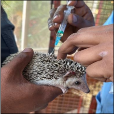
To control the secondary dermatophytosis, the hedgehogs were given a bath with a shampoo containing 2% miconazole and chlorhexidine [5, 6]. The shampoo was lathered on the body, scrubbed with a soft bristle toothbrush, and left on for 10 minutes before being thoroughly rinsed with lukewarm water. Care was taken to avoid letting water enter the eyes, nose, ears, and mouth of the animals. Once dry, selamectin solution @ 45mg/animal [4] (Radicate by Veko) was also applied on the skin in between the spines for all the hedgehogs (Fig. 7). Individual hedgehogs were weighed (Fig. 5) and treated with amoxicillin-clavulanate at 12.5mg/kg BID for 5 days and meloxicam at 0.2mg/kg OD for 3 days, which resolved the dermatitis and aided in the resolution of one hedgehog’s aural haematoma. The animal enclosures were disinfected and sundried to ensure annihilation of ectoparasites and its stragglers [6]
Outcome
The lesions progressively reduced along with the inflammation and swelling, and a stark reduction in itching was observed by day 7 (Fig. 8). Mineral oil ear swab cytology was performed again after 15 days, which was negative for mite infestation. By day 30 of treatment, the hedgehogs were clear of any visible lesions on the body (Fig. 9). Monitoring of the animals was done for 45 days after initial treatment but no signs of re-appearance of symptoms were observed.
Possible causes of infestation:
Otodectes cynotis mites can be transmitted through contact with infected animals or their environments. They can also be transmitted through the use of contaminated bedding or equipment. Poor hygiene and overcrowding may also contribute to the spread of these mites.
Preventive measures
To prevent infestation with Otodectes cynotis mites, it is important to maintain good hygiene and regularly clean and disinfect the hedgehog’s environment. It is also recommended to avoid overcrowding and to avoid sharing bedding or equipment with infected animals. Regular check-ups with a veterinarian can help identify and treat infestations early on.
Conclusion and Discussion
This case report demonstrates the effectiveness of selamectin solution in the treatment of Otodectes cynotis infestation in domestic African pygmy hedgehogs (Fig. 10). Numerous doses are typically required for compounds that have been utilised in the past to treat and manage ectoparasites in exotic pets, which raises the possibility of owner infringement, distressed animal leading to poor therapeutic outcome. Selamectin, an endectocide, does not penetrate the blood brain barrier in the host thereby increasing its margin of safety [7]. It is practical to use against a variety of endo and ecto- parasites because of its lengthy duration of impact and wide spectrum of therapeutic activity.
Dermatophytosis may develop as a result of stresses such as mite infestation. Crusting at the base of the spine is a defining feature of dermatophytosis [6]. This case report also exemplifies the auxiliary use of miconazole and chlorhexidine bath as a remedy against mild secondary dermatophytosis. The use of these treatments resulted in a significant reduction in symptoms and a complete resolution of the mite infestation. It is important to take preventive measures to avoid infestation with Otodectes cynotis mites as acariasis in African pygmy hedgehog is a common finding in individuals subjected to poor husbandry practices and avoiding overcrowding and sharing contaminated bedding or equipment. These findings may be useful for veterinarians treating similar cases in the future.
References
1. Hnilica KA, Patterson AP. Eds., Parasitic Skin Disorders, in Small Animal Dermatology, Fourth. Elsevier; c2017. p. 156. DOI: 10.1016/C2014-0-01191-4.
2. Mitchell MA, Tully TN. Eds., Current therapy in exotic pet practice. St. Louis, Missouri: Elsevier, 2016.
3. Hensel P. Ear Swabs: Technique & Interpretation, Appl. Cytol. 2009, 29-31.
4. Fisher M, Beck W, Hutchinson M, Efficacy and safety of selamectin (Stronghold/Revolution) used off-label in exotic pets. J Appl. Res. Vet. Med. 2007 Jan;5:87-96.
5. Carpenter J, Harms C. Exotic Animal Formulary, Sixth. Missouri: Elsevier, 2023.
6. Meredith AL. British Small Animal Veterinary Association, Eds., BSAVA Manual of exotic pets: a foundation manual, 5. ed. Quedgeley: BSAVA, 2010.
7. Bishop BF, et al. Selamectin: a novel broad-spectrum endectocide for dogs and cats, Vet. Parasitol. 2000 Jul;91(3-4):163-176. Doi: 10.1016/S0304-4017(00)00289-2.
8. Digeronimo P. Therapeutic Review: Selamectin, J Exot. Pet Med. 2015 Nov;25. Doi: 10.1053/j.jepm.2015.11.001.


